Fine Needle Aspiration
For this test, a needle is introduced into the mass, and cells are removed either by sucking them backwards into an empty syringe, or by moving the needle in and out of the lump in different directions several times to collect "cores" of tissue. Then, these cells are placed on a slide, dried and sent off to be viewed by a Board Certified Pathologist; the results usually take about 1 week. Fine needle aspiration is a great, non-invasive technique that can be done easily in most animals without the need for sedation or freezing. It is diagnostic in many types of masses, but can occasionally yield uncertain or non-diagnostic results given the small sample size of cells.
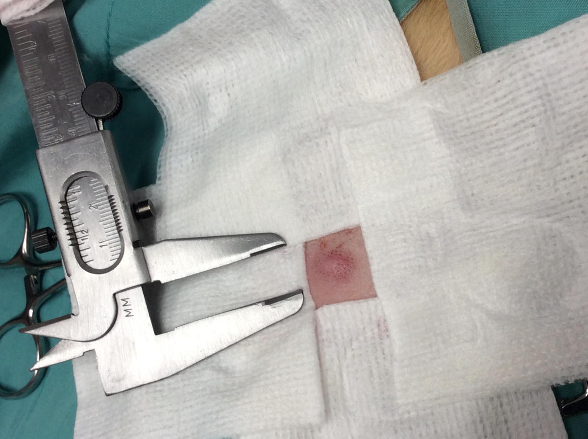
In our patient Mattie Mae's case, the results were surprising. Mattie had a Mast Cell Tumor. Mast Cell Tumors are a common type of malignant tumor found in dogs. They are found in animals both young and old and can have many different appearances making it impossible to diagnose them on appearance alone. They can be aggressive and can spread to other organs. Mattie was taken to surgery and the mass was removed with "wide margins". This is important in aggressive tumors and means we try to remove normal tissue for several centimeters on all sides of the mass. This is one reason why it is important to know what tumor you are removing by diagnosing it as best as possible before surgery. Then we sent the entire mass off to the pathologist. In Mattie's case, it was a "low grade" tumor; meaning, unlikely to have spread. We were successful in removing the entire mass at the surgery.
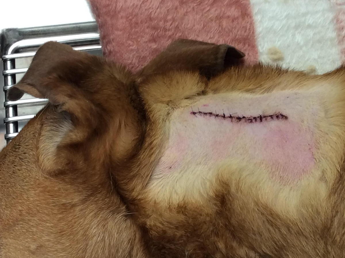
This year, Mattie developed another lump, this time on her neck, and we tested it immediately. It was indeed another Mast Cell Tumor. This did not represent a spread of the first tumor, but rather that she has an increased susceptibility to developing these types of tumors. Again, this one was successfully removed.
Lumps and bumps are a very common concern that we encounter with pets. Sometimes they are the primary reason for a pet to be presented for examination; other times, they are addressed or discovered during routine annual examinations.
Which lumps should we be concerned about?
Firstly, this is one of the reasons why annual examinations are so important! Your veterinarian has the knowledge and experience to determine which lumps we should pay special attention to. The general "rule of thumb" for any mass that is detected, is if it is greater than 1 cm and present for greater than 1 month, it should be examined by your veterinarian.
If your pet has a lump that is GREATER THAN 1 CM FOR GREATER THAN 1 MONTH, book an appointment with your veterinarian to have it examined.
Despite all of our superpowers as veterinarians, we do not have a "microscopic X-ray vision", and we do not have a crystal ball in our pockets that might help us tell you exactly what your dog or cat's lump is made of! Your pet's breed, age, and history of skin tumors or skin disease, as well as palpation of the mass' consistency, location, size, appearance, and rate of growth, will help us form a list of probable causes. However, the true diagnosis can only be obtained with aspiration or biopsy of that mass. With some lumps, we may have a high level of confidence that they are benign and can be monitored, like sebaceous adenomas. Others, like the common "fatty lump", we can have relative confidence, but it is important to understand that more malignant tumors can have an identical appearance.
So this month, Gateway Pet Hospital is launching our inaugural "LUMP MONTH". We encourage you to bring your pet in to have his/her lumps examined, aspirated and sent to a pathologist. Please call us to learn about our "lump month" promotions.