First things first: What is a COHAT?!
COHAT stands for Complete Oral Health Assessment and Treatment. Even with regular home care, routine professional dental cleaning under general anesthesia is an important part of preventative oral care. A professional cleaning is able provide a more thorough cleaning than a toothbrush can achieve. We can get the inside of the teeth and under the gumline where bacteria is most likely to harbour and cause damage. We also have the option to do full mouth dental radiographs (commonly known as x-rays) that can find problems we can't see from the outside! With more advanced disease, extraction of teeth may be required.
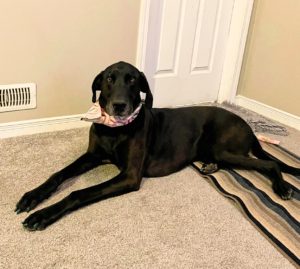
Titus recently underwent a COHAT and his owner (our very own CSR, Sasha) let us document the day so that you can get a better idea of what is happening while your pet is under our care!
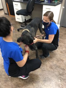
 Prior to an anesthetic procedure we examine Titus to assess for any notable health concerns that could compromise his safety under general anesthesia. We also perform bloodwork to assess for concerns with the liver, kidneys, signs of systemic infection or inflammation, anemia or platelet issues (platelets play a big role in clotting blood which is important for surgery!). Bloodwork, though not required, is recommended for younger patients. However, as our pets age, the likelihood of underlying issues increases so we do require this before elective procedures in patients over the age of 6.
 Titus is then given an injection of a sedative and pain medication, typically this concoction is referred to as a "premedication". The dose and type of medication used is tailored to each patient. These medications allow us to use lower doses of anesthesia, which, in turn, makes the anesthetic safer.
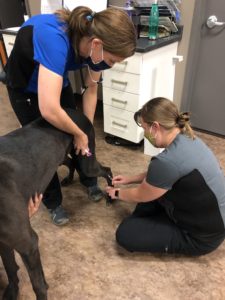
A catheter is then placed in Titus' leg. A catheter is an extremely important part of general anesthesia. It not only allows intravenous fluids to be delivered to help support Titus' blood pressure but also allows emergency intravenous access if Titus was to experience any complications.
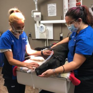
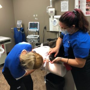
Induction of anesthesia is done by injection into the catheter. We inject an initial dose of anesthesia and then assess Titus. Some animals require higher doses than others and so we titrate (give small amounts to effect) to avoid giving more than necessary. Once Titus is completely unconscious an endotracheal tube is placed. This tube keeps Titus' airway open and allows us to deliver oxygen and inhalant anesthesia. It also protects the airway in case of regurgitation, which is when the stomach contents come up and prevents those contents from going down the trachea and into the lungs. The risk of regurgitation is also why Titus was fasted before coming in for his dental procedure.
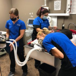
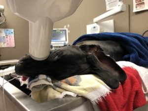
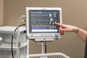 A Registered Veterinary Technician (RVT) trained in anesthesia monitors Titus' vital signs throughout the entire procedure. We monitor his heart rate and electrical activity with an ECG, his oxygen saturation with a pulse oximeter, his respiration rate and carbon dioxide levels with a CO2 monitor as well as his blood pressure. These machines are extremely helpful tools but aren't worth much without the skill and expertise of the veterinary technician and veterinarian to read and interpret what the machines are telling us and to act accordingly if his vital signs were to indicate any concerns.
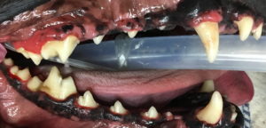
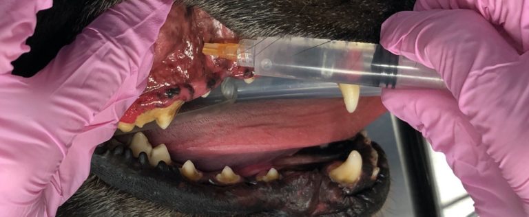
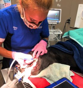 Titus's teeth are cleaned by a Registered Veterinary Technician. First, an ultrasonic scaler is used to clean all the surfaces of the teeth. While brushing at home can be helpful to slow down the development of plaque and tartar, it is nearly impossible to clean the inside surfaces or the teeth or those further back in the mouth. Once the teeth have been cleaned the polisher is used to smooth out any microscopic grooves made by the scaler.
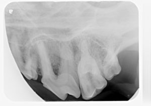
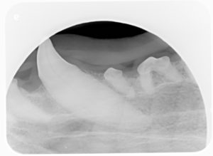
Dental radiographs are taken to evaluate the roots of the teeth. Teeth are like icebergs, we can only see a portion of the tooth above the gumline. Radiographs are an extremely valuable tool to ensure we are not missing sources of pain in our pet's mouths. We also evaluate each tooth with a dental probe to assess for any periodontal disease. With the combination of the visual exam of the mouth and evaluation of the dental radiographs we then determine if any additional treatment is required beyond a dental cleaning. Two of Titus' radiographs are shown below - these radiographs show nice healthy teeth!
In Titus' case he did not require any dental extractions and so was recovered at this stage. If we had identified any pathology that required the extraction of teeth we would proceed to the next steps. We have used photos from various other patients for the extraction portion of this post!
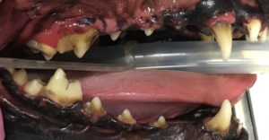
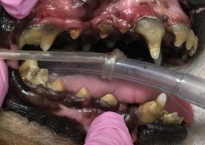
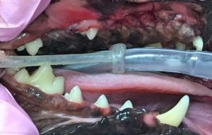
 (Titus’ before and after photos are shown here. He has some enamel staining that cannot be removed with cleaning however this staining is only of cosmetic concern and not harmful to Titus.)
Prior to dental extractions we place a nerve block (freezing). This increases anesthetic safety as the absence of pain at the surgical site means that lower doses of anesthetic is needed to keep the patient unconscious throughout the procedure. Sometimes we will even do an IV drip of pain medications to help ensure our patient stays comfortable and our anesthetic doses can stay low.
All oral surgery, including extractions, are performed by the veterinarian. The technique used depends on the tooth in question as some are single rooted and others are multi-rooted. In general the process includes loosening the gums to gain access to the bone covering the tooth, the use of a drill to remove some of the bone and then specific dental tools to loosen the ligaments holding the tooth in place and ultimately remove the tooth. We then suture (stitch) the gums closed. Unlike in human dentistry, where we can ensure to keep the defect left behind clean using a syringe and water, this is very difficult in our pets and so closure of the site is important. The sutures used will dissolve in a few weeks.
 (This patient had advanced periodontal disease that required numerous extractions. You can see the teeth that have been extracted and sutured closed. This patient likely felt so much better after having these sources of chronic pain removed!!!)
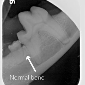
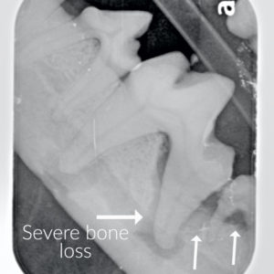 (These are dental radiographs from different patients. The radiograph on the left shows normal teeth and bone. The radiograph on the right there is a deep periodontal pocket and bone loss associated with two of the teeth. These pockets and bone loss will only worsen. In most cases the tooth will loosen over time and fall out, though painfully. However in rare cases the jaw could fracture as a result of bone loss. Extraction is the recommended treatment for these teeth.)
Once the dental procedure is complete, the patient is carefully recovered by the RVT. As the patient awakens from anesthesia, the endotracheal tube is removed. The timing of this is important to ensure that the patient is conscious enough to control their own breathing and swallow reflexes before the tube is removed. We also need to ensure that the patient remains calm upon recovery and that their pain levels are well controlled. Medications are prepared ahead of time and are on-hand should they be needed.
The patient is monitored in hospital for several hours postoperatively and once they are fully recovered they will be discharged with instructions for their care and pain medication if extractions have been performed. In Titus' case, because no extractions were performed, an injection of an anti-inflammatory is given during his procedure and provides sufficient pain control. No diet change is required. However, in cases that require extraction the patient will need to be fed soft food while the sites heal (approximately 2 weeks) and will be sent home with additional pain control.